Sickle Cell Anemia Investigation: Day 5
Today did not go too well. I must remind myself that it is quite a lot for students to jump from DNA to mRNA to Protein Shape & Function to Cell Shape to bodily function. Still, I think today should have worked much better than it did.
We started with a video by BioVisions at Harvard titled Cellular Visions. It shows how many different types of proteins function in a cell.
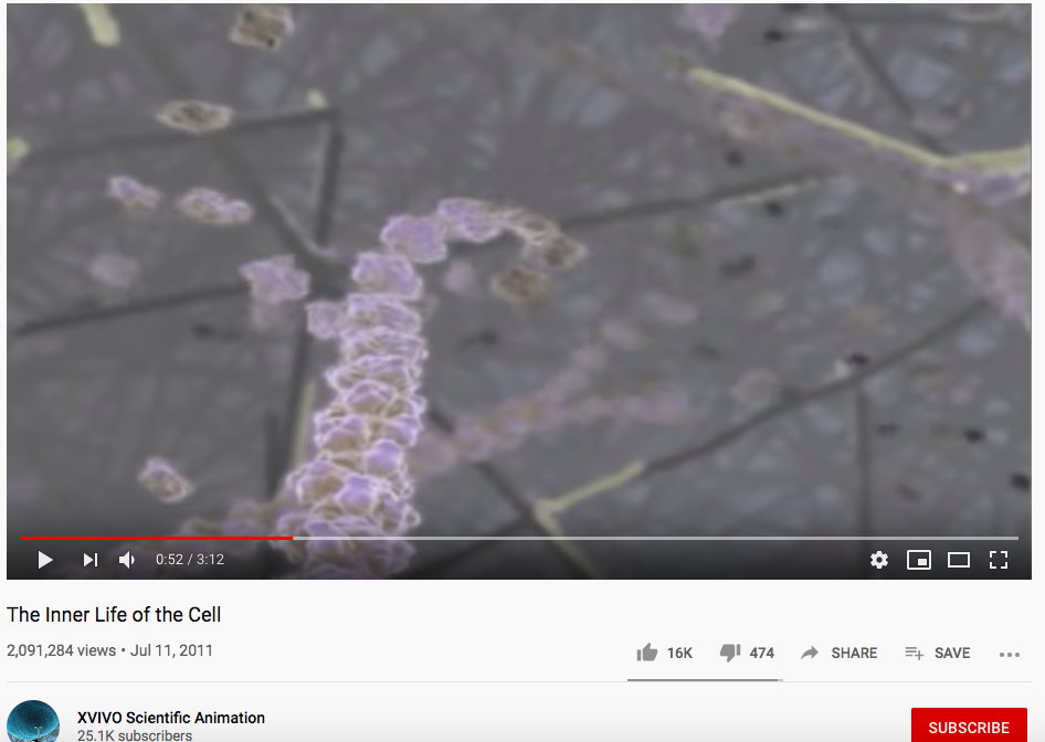
Then we reviewed that cells shape helps them to perform their function and that their shape is determined by a sequence of amino acids. I told several other protein ‘stories’. I then ended with a visual from Utah Genetics that shows cell size and scale so that I could compare a red blood cell and a hemoglobin protein. I still have many students who cannot interpret that cells are much larger than proteins.

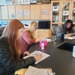
Next, I tried to do a hands on activity with the students that I got from HHMI titled How Do Fibers Form. I think the lesson should work but I couldn’t get it to click. I had lots of blinking and phone issues instead of engaged hands and minds.
Essentially, students take cut out paper models of hemoglobin and they try to piece them in a cell to see how the proteins interact and determine the shape of the cell.
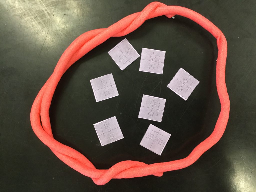
I pulled up David Goodsell’s Molecule of the month blog to show the students the behavior of this hemoglobin molecule.
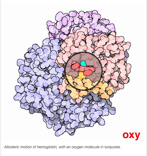
On the other hand when an oxygen is not bound to the heme group of hemoglobin it enters an “open” configuration.
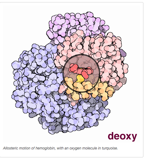
We can model this with a piece of paper that has a hole punch to show that it is open.

I ask the students to show how these proteins might interact if they were bouncing around a cell. None of my students got this at all…There were literally crickets chirping in my Leopard Gecko cage folks… pppsssssssth 🙁
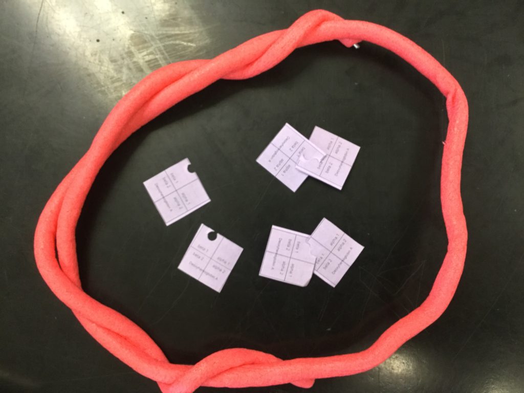
Next, I had them manipulate models of hemoglobin that had been mutated.
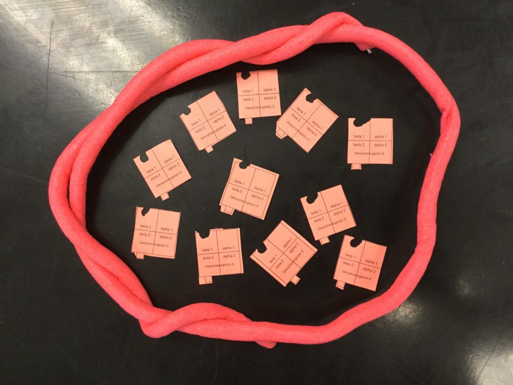
Actually, several of the students started stringing them together… but none (literally 0/111) of them put together that this growing chain of proteins could actually push on the cell membrane to change the shape.
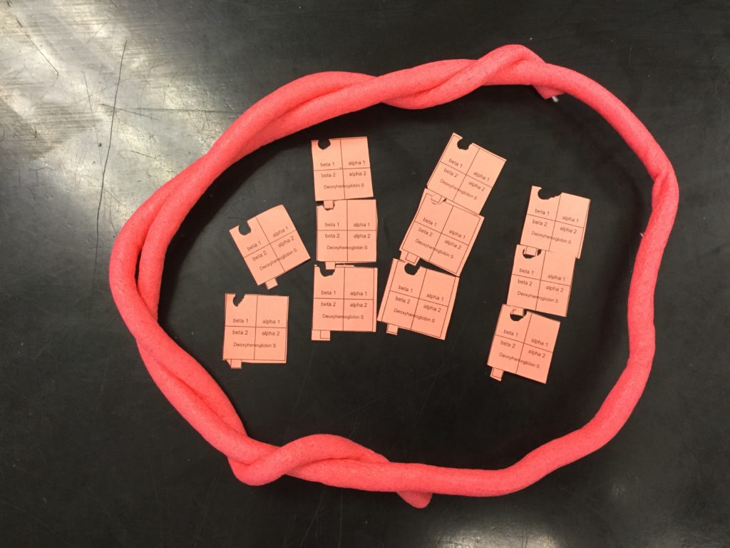
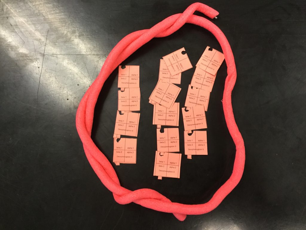
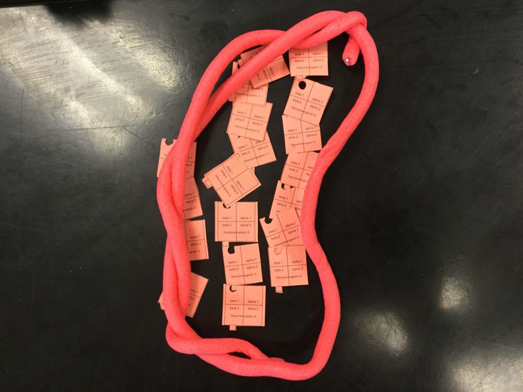
I actually did a clearer example with better models that I had printed off.
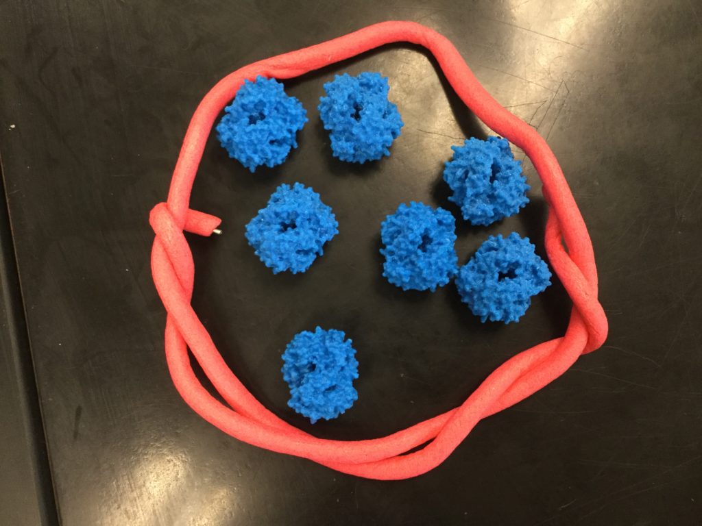
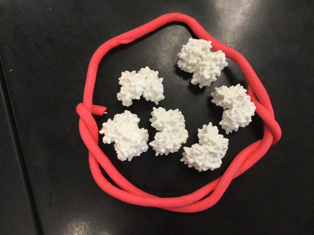
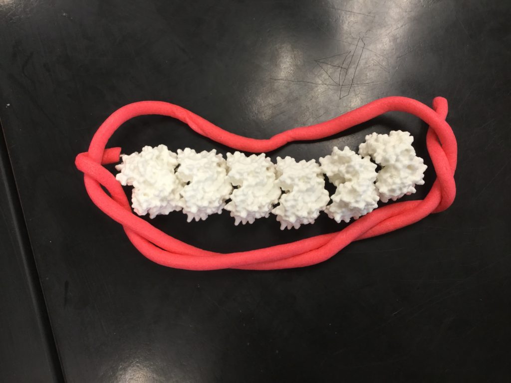
By the last hour I simply just showed them how this could work with my overhead projector. Then we went outside with the last 10 minutes of class and played hacky sack.
I am going to try again on Monday.
 Next Post